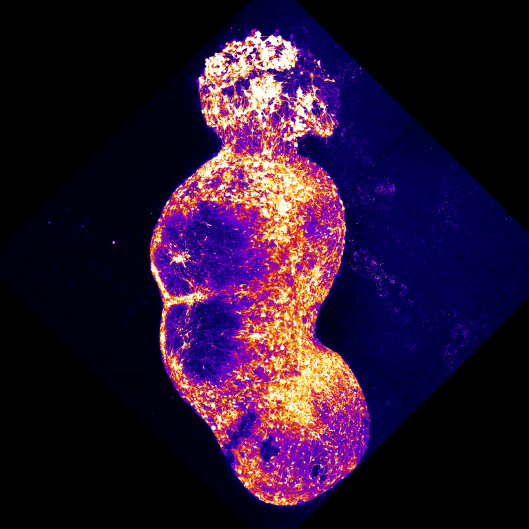
Building a human sensory relay
Four connected human stem-cell models let researchers watch sensory signals travel from peripheral neurons toward the cortex—and ask how pain-associated mutations change the whole circuit.
Read the article →An experimental guide to the measurement, the biological question, and the limits of interpretation.
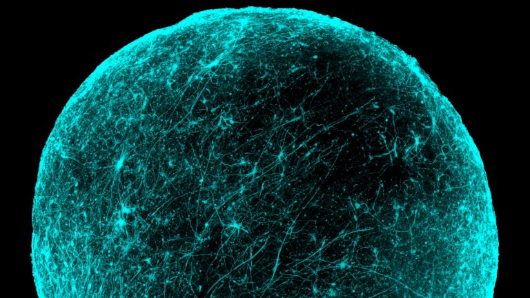
Three-dimensional cell models reproduce selected features of tissues under laboratory conditions. Organoids and region-specific spheroids allow researchers to study cellular organization and development. Assembled models bring distinct tissue components together to investigate interactions that a single component cannot reproduce.
In the Stanford-led forebrain spheroid study, researchers joined dorsal-like and ventral-like tissues derived from human pluripotent stem cells. They measured neuronal migration with live imaging and tested electrical communication after integration. Such readouts connect a model’s structure with specific functions. Comparing donor-derived tissues or changing experimental conditions can help test mechanisms, provided the model is characterized and the measured event is clearly defined.
An organoid is most useful when the question is precise: how do particular cells differentiate, migrate, or interact? Region-specific models can isolate a developmental process. Assembloids combine components to study interactions between them. Choosing what to include also determines which biological processes remain outside the model.
Gene-expression profiles and protein markers help establish which cells are present. Functional measurements ask what those cells actually do. In neural models, live imaging can measure migration while electrical recordings probe communication. A model’s three-dimensional shape alone does not establish that it reproduces the function of a mature organ.
Different donor lines and batches can produce different cellular mixtures and developmental timing. Strong comparisons account for those sources of variation and specify the independent biological units. A rescue of a cellular phenotype can test a mechanism without proving that the same intervention would improve symptoms in a person.
These models represent selected tissue features and lack the full anatomy, blood supply and environment of a living organ.
Four connected human stem-cell models let researchers watch sensory signals travel from peripheral neurons toward the cortex—and ask how pain-associated mutations change the whole circuit.
Read the article →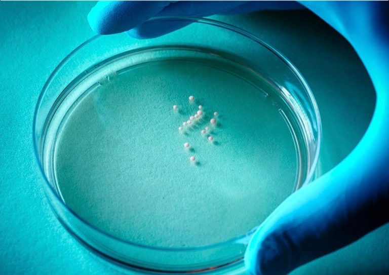
Patient-derived organoids and assembloids test an antisense strategy that reduces a disease-associated calcium-channel exon without removing the channel itself.
Read the article →Human forebrain spheroids made a hidden developmental process visible: neurons moving into a new region and joining its circuits.
Read the article →